 Return to the tutorial menu.
General FeaturesOsteoporosis is accelerated bone loss. Normally, there is loss of bone mass with aging, perhaps 0.7% per year in adults. However, bone loss is greater in women past menopause than in men of the same age. The process of bone remodelling from resorption to matrix synthesis to mineralization normally takes about 8 months--a slow but constant process. Bone in older persons just isn't as efficient as bone in younger persons at maintaining itself--there is decreased activity of osteoblasts and decreased production of growth factors and bone matrix. 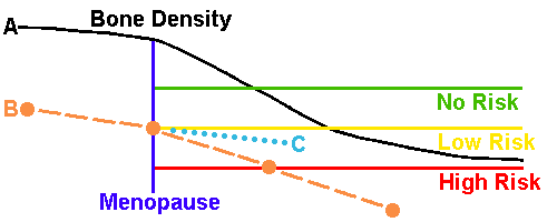
This diagram illustrates changes in bone density with aging in women. The normal curve (A) steepens following menopause, but even by old age the risk for fracture is still low. A woman who begins with diminished bone density (B) even before menopause is at great risk, particularly with a more accelerated rate of bone loss. Interventions such as postmenopausal estrogen (with progesterone) therapy, the use of drugs such as the non-hormonal compound alendronate that diminishes osteoclast activity, and the use of diet and exercise regimens can help to slow bone loss (C) but will not stop bone loss completely or restore prior bone density. Diet and exercise have a great benefit in younger women to help build up bone density and provide a greater reserve against bone loss wiht aging. Risk factors for osteoporosis include:
Osteoporosis can be classified as primary or secondary. Primary osteoporosis is simply the form seen in older persons and women past menopause in which bone loss is accelerated over that predicted for age and sex. Secondary osteoporosis results from a variety of identifiable conditions that may include:
Modifiable risk factors that may potentiate osteoporosis include:
DiagnosisDiagnosis of osteoporosis is made by three methods:
Of these three the best is radiographic bone density measurement. A variety of techniques are available, including single-photon absorptiometry, dual-photon absorptiometry, quantitative computed tomography, dual x-ray absorptiometry, and ultrasonography. Most often, site specific measurements are performed. The most common sites analyzed are those with greatest risk for fracture: hip, wrist, and vertebrae. The forearm and heel that are easily measured using single-photon absorptiometry, quantitative computed tomography, and ultrasonography can be inexpensive, but these sites are typically unresponsive to therapy and give less information about response to therapy. Increased risk for fracture correlates with decreasing bone density. Serial measurements over time can also give an indication of the rate of bone loss and prognosis. The two main biochemical markers for bone formation are serum alkaline phosphatase and serum osteocalcin. Markers for bone resorbtion include urinary calcium and urinary hydroxyproline:
Bone biopsy is not often utilized for assessment of bone density. This test has limited availability, and is best utilized as a research technique for analysis of treatment regimens for bone diseases. The best clinical use of bone biopsy combines double tetracycline labelling to determine appositional bone growth and rule out osteomalacia. Doses of tetracycline are given weeks apart, and the bone biopsy is embedded in a plastic compound, sliced thinly, and examined under fluorescent light, where the lines of tetracycline (which autofluoresce) will appear and appositional growth assessed. Consequences of OsteoporosisOsteoporotic bone is histologically normal in its composition--there is just less bone. This results in weakened bones that are more prone to fractures with trauma, even minor trauma. The areas most affected are:
Hip fractures that occur, even with minor falls, can be disabling and confine an elderly person to a wheelchair. It is also possible to surgically put in a prosthetic hip joint. Wrist fractures are common with falls forward with arms extended to break the fall, but the wrist bones break too. Vertebral fractures are of the compressed variety and may be more subtle. Vertebral fractures may result in back pain. Another consequence is shortening or kyphosis (bending over) of the spine. This can lead to the appearance of a "hunched over" appearance that, if severe enough, can even compromise respiratory function because the thorax is reduced in size. Persons suffering fractures are at greater risk for death, not directly from the fracture, but from the complications that come from hospitalization with immobilization, such as pulmonary thromboembolism and pneumonia. Osteoporosis is so common that, on average, about 1 in 2 elderly Caucasian women will have had a fracture. In contrast, only about 1 in 40 men of similar age will have had a fracture. Men start out with a greater bone mass to begin with, so they have a greater reserve against loss. Prevention StrategiesThe best long-term approach to osteoporosis is prevention. If children and young adults, particularly women, have a good diet (with enough calcium and vitamin D) and get plenty of exercise, then they will build up and maintain bone mass. This will provide a good reserve against bone loss later in life. Exercise places stress on bones that builds up bone mass, particularly skeletal loading from muscle contraction with weight training exercises. However, any exercise of any type is better than none at all, and exercise also provides benefits for prevention of cardiovascular diseases that are more common in the elderly. Athletes tend to have greater bone mass than non-athletes. Exercise in later life will help to retard the rate of bone loss. TreatmentPersons with osteoporosis may benefit from an improved diet, including supplementation with vitamin D and calcium, and moderate exercise to help slow further bone loss. Most drug therapies work by decreasing bone resorbtion. At any given time, there is bone that has been resorbed but not replaced, and this accounts for about 5 to 10% of bone mass. By decreasing resorbtion of bone, a gain in bone density of 5 to 10% is possible, taking about 2 to 3 years. However, no drug therapy will restore bone mass to normal. Women past menopause with accelerated bone loss may benefit from hormonal therapy using estrogen with progesterone. The estrogen retards bone resorption and thus diminishes bone loss. This effect is most prominent in the first years after menopause. One of the more common non-estrogen therapies is the use of biphosphonates such as alendronate or risedronate that act an an inhibitor of osteoclastic activity. Biphosphonates may be beneficial, particularly in women who cannot tolerate estrogen therapy. Biphosphonaes are effective in inhibiting bone loss after menopause. In one study risedronate has shown effectiveness in reducing the risk of hip fracture among elderly women with osteoporosis. Raloxifene is a selective estrogen receptor modulator (SERM) that may also replace estrogen therapy. Raloxifene can act in concert with estrogen in bone to inhibit resorbtion and decrease the risk for fractures. Though raloxifene inhibits bone resorbtion, it does not have an anabolic effect. Additional potential benefits from raloxifene therapy include decreased risk for breast cancer, because raloxifene acts antagonistically to estrogen on the uterus. Conversely, raloxifene acts in concert with estrogen to protect against and reduce atherogenesis. Teriparatide is a recombinant human parathyroid hormone administered by subcutaneous injection which binds to specific high-affinity cell-surface receptors in bone and kidney, similar to the 34 N-terminal amino acids of parathyroid hormone, and has the same physiological actions on bone and kidney. Daily administration of teriparatide stimulates new bone formation by promoting osteoblastic activity over osteoclastic activity, improving trabecular bone architectural remodelling and increasomg bone mass. Other drug therapies are less commonly employed. Calcitonin, a hormone that decreases bone resorbtion, may be taken by injection or by nasal spray. Sodium fluoride can increase the measured bone density in vertebra, but seems to have no overall effectiveness in reducing vertebral fracture. Examples of Osteoporosis
References
|