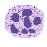
White Cell Differential Count
A manual WBC differential count is performed by having a person trained in peripheral blood morphology review the stained blood smear and manually count 100 white cells (or 50 cells in the case of severe leukopenia). The major advantage is that the observer can determine subtle differences in morphology and observe additional changes in RBC morphology and platelets. The major disadvantage is the need for a trained person to spend increased time (with increased cost) needed to scan the smears. Also, there can be some inter-observer variation.
A standard complete blood count is performed on an automated laboratory instrument that quantitates the number of WBC's present. Some instruments are also able to perform an "automated" WBC count. The advantages of the automated WBC differential are speed, low cost per test, and the precision from the large number of cells counted. The disadvantages include the initial cost of buying the instrument, inability to distinguish subtle differences in morphology, more marked abnormalities, and lack of information about RBC's or platelets.
An example of a CBC with automated WBC differential count is shown below. The absolute numbers for total WBC count and each type of leukocyte are in thousands per cubic millimeter (or alternatively, per microliter).

The absolute white blood cell counts are most useful, and are calculated by multiplying the % of each cell type counted by the total WBC count. Simple percentages may be misleading, since an apparent percentage increase in one constituent may actually be due to a significant and absolute decrease of another type of WBC. For example, 99% lymphs with an absolute WBC count of 1300/microliter means neutropenia, not lymphocytosis. Always consider the WBC percentages in the context of the total WBC count.
Nomenclature
The following terms are used in describing the morphology of WBC's, as seen on a standard peripheral blood smear:
- Neutrophilia
An increase in the absolute neutrophil count, it can be increased transiently with stress and exercise by a shift of neutrophils from the marginating pool to the circulating pool. Pathologic processes that result in neutrophilia include:
Infection
Toxins: metabolic (uremia), drugs, chemicals
Tissue destruction or necrosis: infarction, burns, neoplasia, etc
Hemorrhage, especially into a body cavity
Rapid hemolysis
Hematologic disorders: leukemias, myeloproliferative disorders
- Neutropenia
A decrease in the absolute neutrophil count. Pathologic processes that result in neutropenia include processes that decrease production or increase destruction. Diseases that decrease neutrophil production include:
Aplastic anemia
Toxins that damage marrow
Collagen vascular diseases (such as SLE)
Myelphthisic marrow processes such as marrow infiltration by infections or metastatic carcinomas
Hematologic malignancies such as leukemias
Myeloproliferative disorders
Radiation therapy
Chemotherapy
Congenital disorders
Diseases that increase neutrophil destruction include:
Splenomegaly with hypersplenism
Infection
Immune destruction
- Lymphocytosis
An increase in the number of circulating lymphocytes may normally be observed in infants and young children. Pathologic processes with lymphocytosis may include:
Acute infections, including pertussis, typhoid, and paratyphoid
Infectious mononucleosis, with "atypical" lymphocytosis
Viral infections, including measles, mumps, adenovirus, enterovirus, and Coxsackie virus
Toxoplasmosis
HTLV I
- Lymphopenia
A decrease in the number of circulating monocytes may be seen with:
Immunodeficiency syndromes, including congenital (DiGeorge syndrome, etc) and acquired (AIDS) conditions
Corticosteroid therapy
Neoplasia, including Hodgkin's disease, non-Hodgkin's lymphomas, and advanced carcinomas
Radiation therapy
Chemotherapy
- Monocytosis
An increase in the number of circulating monocytes may be seen with:
Infections: such as brucellosis, tuberculosis and rickettsia
Myeloproliferative disorders
Hodgkin's disease
Gastrointestinal disorders, including inflammatory bowel diseases and sprue
- Monocytopenia
A decrease in the number of circulating monocytes may be seen with:
Early corticosteroid therapy
Hairy cell leukemia
- Eosinophilia
An absolute increase in the number of circulating eosinophils may occur with:
Allergic drug reactions
Parasitic infestations, especially those with tissue invasion
Extrinsic asthma
Hay fever
Extrinsic allergic alveolitis ("farmer's lung"
Chronic infections
Hematologic malignancies: CML, Hodgkin's disease
- Eosinopenia
An absolute decrease in the number of circulating eosinophils may occur with:
Acute stress reactions with increased glucocorticoid and epinephrine secretion
Acute inflammation
Cushing's syndrome with corticosteroid therapy
- Basophilia and Basopenia
An absolute increase in the number of circulating basophils may occur with myeloproliferative disorders and with some allergic reactions.
An absolute decrease in the number of circulating basophils may occur with the same conditions that lead to eosinopenia.
Morphologic Findings
The following terms are used in describing the morphologic variation seen in WBC's on a standard peripheral blood smear:
|
 Return to the tutorial menu.


