 Return to the radiologic techniques menu.
Return to the radiologic techniques menu.
Magnetic resonance imaging (MRI), or magnetic resonance (MR) scanning, is done with a large magnet and pulsed radiofrequency waves. The magnet is so powerful that large metallic objects such as pacemakers cannot be put into the MR scanner.
The magnet aligns the hydrogen atoms (themselves tiny magnets with a single proton) within tissues. Each proton acts like a tiny spinning gyroscope. It is the water in tissues that contributes the most to this effect, since each water molecule has two hydrogens atoms bound with one oxygen atom (H2O). As the hydrogen atoms are aligned, pulsed radiofrequency (RF) waves are directed at the hydrogen atoms, which disturb their alignment. They shift alignment, much as square dancers respond to the music and the caller. As they realign, they emit the absorbed radiofrequency energy, which is detected and converted to a signal that is analyzed by a computer to create an image.
There are several modes of MR scanning, based upon how long it takes the hydrogen atoms to realign, called the "relaxation time." The "longitudinal" relaxation time is called T1, while the "transverse" relaxation time produces a T2 weighted scan.
The intensity of the MR signal depends upon the scanning mode (e.g., T1) and the type of tissue being scanned. The tissues with high signal intensity with T1 scans include those with adipose tissue--fat appears very bright--while bone appears black. With a T2 weighted scan, fat is not as bright, but fluids are very bright.
One major advantage to MRI is that images can be generated in multiple planes from a single scan. The computer can generated axial, coronal, and sagittal images. The images can be generated after the scan is complete, because the data from the scan are stored in the computer. A scan of a body region may take more than 30 minutes. Thus, movement is more likely to produce artefact in MR scans than in CT scans.
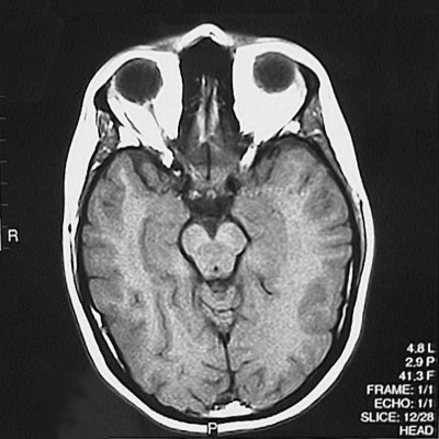
The regions most amenable to MRI are brain and extremities, which have no movement (with patient cooperation and/or sedation). Chest and abdominal MR scans can be done, but will not be as sharp as CT images. Sometimes a gadolinium contrast material is administered intravenously to the patient undergoing brain MR scanning; this helps to highlight abnormalities such as neoplasms. An axial MR T1 weighted scan section of a normal brain is seen below. Note that the skull bone is black, while the orbital fat is bright white.
An axial MR T2 weighted scan section of a normal brain is seen below. Note that the cerebrospinal fluid in the subarachnoid spaces and the vitreous humor of the eyes is now bright.
