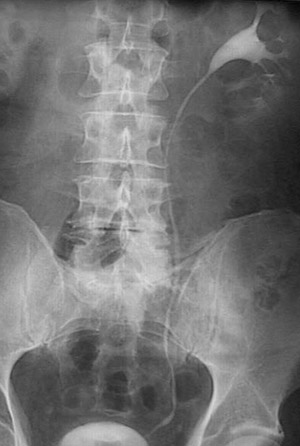
This intravenous pyelogram (IVP) of a normal urinary tract on the left demonstrates contrast material that is filling the
pelvis
, then the
ureter
, and finally the
bladder
.