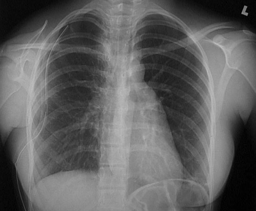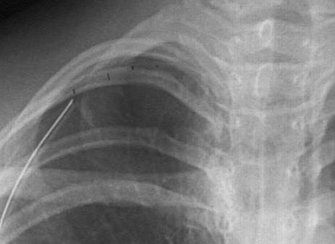


| The radiograph above demonstrates a pneumothorax on the right, with the mediastinum and heart shifted to the left. A pneumothorax results from rupture of the lung or a penetrating injury to the chest wall that allows air to enter the pleural cavity. A chest tube has been inserted here to help re-expand the lung. The faint border of the displaced visceral pleura surface is marked in the radiograph below. |


 |