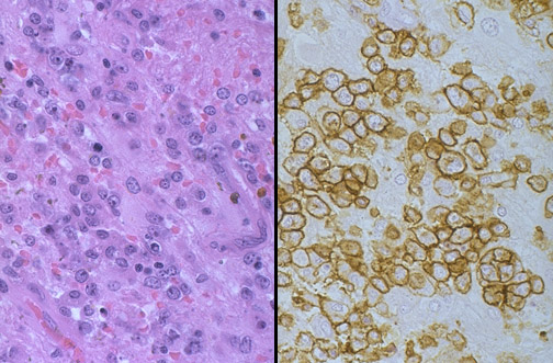
The CNS neoplasm seen with H&E staining on the left is poorly differentiated, so the immunoperoxidase stain on the right with antibody to CD-45, or "common leukocyte antigen" that stains lymphoid cells, helps to identify the neoplasm as a lymphoma.