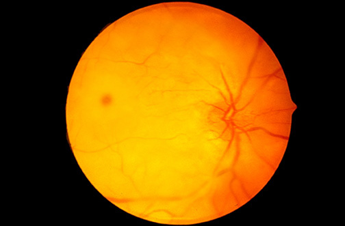


| There is paleness to the retina, with a "cherry red" spot, which can be a feature of retinal arterial occlusion or of Tay-Sachs disease. The extensive opacification here is more characteristic for Tay-Sachs disease, which results from a deficiency in hexosaminidase A enzyme with accumulation of GM2 trihexosylceramide in retinal ganglion cells, producing the pale appearance that obscures the vascularity. The greatest density of ganglion cells in the macular area leads to greater opacification, except in the foveal pit that is devoid of ganglion cells, so that a "cherry red" spot is seen. The lipids accumulating in retinal ganglion cells lead to ganglion cell hypertrophy, followed by cell death and eventual gliosis and blindness. |


 |