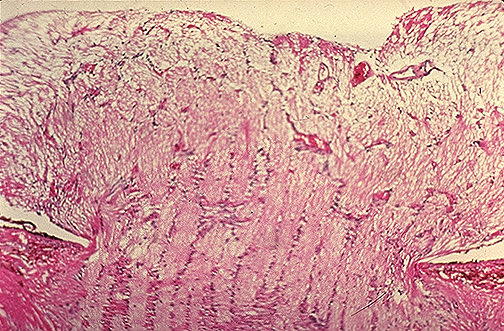


| This microscopic section through the head of the optic nerve displays papilledema. Note the bulging of the nerve above the level of the surrounding retina with forward bowing of the lamina cribrosa. The intracranial pressure must be relieved, or the patient may suffer herniation (cerebellar tonsils, uncus of hippocampus, cingulate gyrus). [Image contributed by Nick Mamalis, MD, University of Utah] |


 |